| dc.contributor.author | Fabre, Stéphanie | - |
| dc.contributor.author | Lang, Valérie | - |
| dc.contributor.author | Bismuth, Georges | - |
| dc.date.accessioned | 2014-07-03T06:52:31Z | |
| dc.date.available | 2014-07-03T06:52:31Z | |
| dc.date.issued | 2006 | fr_FR |
| dc.identifier.citation | Fabre, Stéphanie ; Lang, Valérie ; Bismuth, Georges ; La PI3-kinase : de la synapse immunologique au contrôle de la prolifération T, Med Sci (Paris), 2006, Vol. 22, N° 10; p. 872-877 ; DOI : 10.1051/medsci/20062210872 | fr_FR |
| dc.identifier.issn | 1958-5381 | fr_FR |
| dc.identifier.uri | http://hdl.handle.net/10608/5881 | |
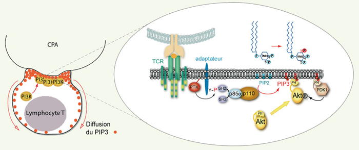
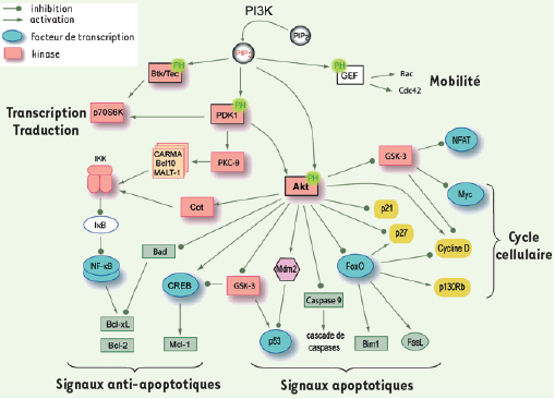
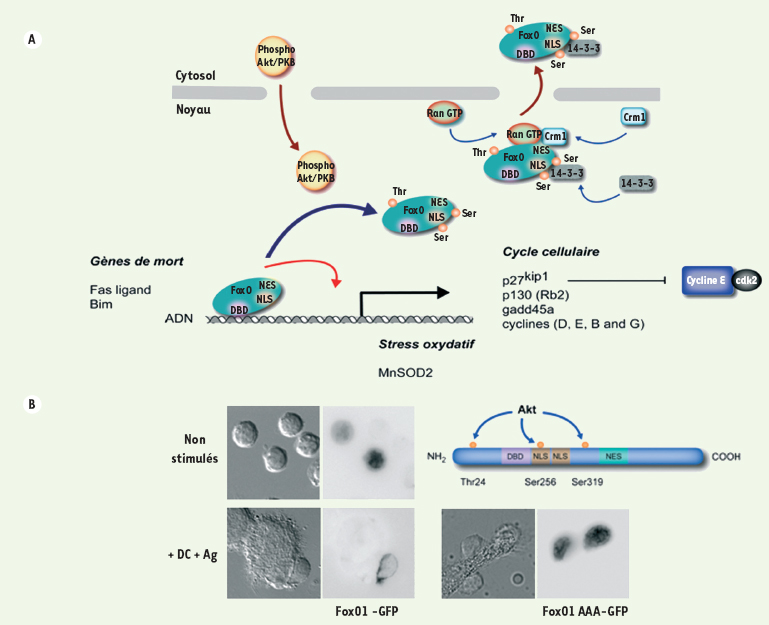
| dc.description.abstract | Le contrôle de la quiescence lymphocytaire est un élément essentiel à l’homéostasie du système immunitaire. Cet état quiescent est activement maintenu par des facteurs transcriptionnels nucléaires, de la famille FoxO (Forkhead subgroup O) notamment. Cet équilibre est cependant rompu à la suite de la reconnaissance de l’antigène, ce qui déclenche la division cellulaire. Cet article fait le point des données récemment acquises sur les mécanismes moléculaires impliqués dans ce phénomène. Il décrit ainsi comment la synapse immunologique formée entre un lymphocyte T et une cellule présentant l’antigène constitue une plate-forme d’intégration au sein de laquelle il existe une production continue de 3’-phospho-inositides sous l’impulsion des phosphoinositides-3-kinases. En provoquant l’activation de la sérine-thréonine kinase Akt, ce processus stimule l’exclusion nucléaire des FoxO pour lever le frein permanent qu’ils exercent et permettre l’expansion clonale des cellules T en réponse à l’antigène. | fr |
| dc.description.abstract | T cell clonal expansion contributing to host defense against pathogens is a tightly controlled process to maintain the homeostasis of the immune system. Our understanding of how T cell growth and proliferation are controlled following antigenic stimulation is therefore a major challenge. Antigen recognition occurs when a naive T lymphocyte contacts an antigen-presenting cell. A specialized junction enriched in T-cell receptors, costimulation molecules and signaling adaptors, called the immunological synapse, is then created for several hours between the two cell types. Recent discoveries now clarify the molecular mechanisms used by this organization to control T cell growth and proliferation. It has been established that the immunological synapse functions in fact as an integrative platform where class Ia phosphoinositide-3-kinases (PI3Ks) are recruited and activated to continuously produce high levels of 3’-phosphoinositides. These lipids regulate the localization and the activation of a wide range of PH-domain containing proteins, among which the serine-threonine kinase Akt, a downstream effector of PI3Ks, appears to be a key player. FoxO (Forkhead subgroup O) family members control in various cell systems genes implicated in apoptosis, stress resistance and cell cycle arrest, thereby contributing to maintain quiescence in unstimulated cells. In naïve T cells contacting antigenpresenting cells a rapid but also very prolonged nuclear exclusion of these transcription factors is observed downstream of Akt. Mainly, this compartmentalization process is mandatory to induce T cell growth triggered findings demonstrate that to initiate cell cycle progression the formation of the immunological synapse is an undemanding tactic used by primary T cells to securely maintain the 3’-phosphoinositide-dependent mitotic switch governed by the spatial control of FoxO transcription factors. | en |
| dc.language.iso | fr | fr_FR |
| dc.publisher | EDK | fr_FR |
| dc.relation.ispartof | M/S revues | fr_FR |
| dc.rights | Article en libre accès | fr |
| dc.rights | Médecine/Sciences - Inserm - SRMS | fr |
| dc.source | M/S. Médecine sciences [ISSN papier : 0767-0974 ; ISSN numérique : 1958-5381], 2006, Vol. 22, N° 10; p. 872-877 | fr_FR |
| dc.subject.mesh | Prolifération cellulaire | fr |
| dc.subject.mesh | Phosphatidylinositol 3-kinases | fr |
| dc.subject.mesh | Synapses | fr |
| dc.subject.mesh | Lymphocytes T | fr |
| dc.title | La PI3-kinase : de la synapse immunologique au contrôle de la prolifération T | fr |
| dc.type | Article | fr_FR |
| dc.contributor.affiliation | Inserm U567, CNRS UMR 8104, Université Paris 5, Faculté de Médecine René Descartes, Institut Cochin, Département de Biologie cellulaire, 22, rue Méchain, 75014 Paris, France | fr_FR |
| dc.identifier.doi | 10.1051/medsci/20062210872 | fr_FR |
| dc.identifier.pmid | 17026942 | fr_FR |