| dc.contributor.author | Félétou, Michel | - |
| dc.contributor.author | Busse, Rudi | - |
| dc.contributor.author | Edwards, Gillian | - |
| dc.contributor.author | Fleming, Ingrid | - |
| dc.contributor.author | Weston, Arthur H. | - |
| dc.contributor.author | Vanhoutte, Paul M. | - |
| dc.date.accessioned | 2014-01-09T10:56:30Z | |
| dc.date.available | 2014-01-09T10:56:30Z | |
| dc.date.issued | 2003-12 | fr_FR |
| dc.identifier.citation | Félétou, Michel ; Busse, Rudi ; Edwards, Gillian ; Fleming, Ingrid ; Weston, Arthur H. ; Vanhoutte, Paul M. ; Dialogue entre cellules endothéliales et cellules musculaires lisses, Med Sci (Paris), 2003, Vol. 19, N° 12; p. 1242-1250 ; DOI : 10.1051/medsci/200319121242 | fr_FR |
| dc.identifier.issn | 1958-5381 | fr_FR |
| dc.identifier.uri | http://hdl.handle.net/10608/4660 | |
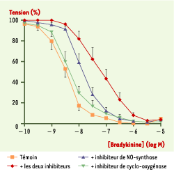
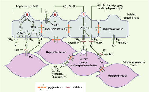
| dc.description.abstract | Les cellules endothéliales sont des cellules qui jouent, entre autres, un rôle actif et essentiel dans le contrôle du tonus vasculaire, et donc du débit sanguin local, en sécrétant divers agents vasoconstricteurs (endothéline, prostaglandines) ou vasodilatateurs (prostacycline, monoxyde d’azote). Un mécanisme vasodilatateur supplémentaire, associé à une hyperpolarisation des cellules musculaires lisses, est observé principalement dans la circulation coronaire et dans les lits vasculaires périphériques. Ce phénomène, attribué à un facteur élusif dénommé facteur hyperpolarisant dépendant de l’endothélium (EDHF, endothelium-derived hyperpolarizing factor), est maintenant partiellement élucidé : il implique une augmentation du calcium intracellulaire, suivie d’une hyperpolarisation de la cellule endothéliale grâce à l’ouverture de canaux potassiques dépendant du calcium. L’hyperpolarisation des cellules endothéliales est alors transmise par diverses voies aux cellules musculaires lisses et se propage le long de l’axe vasculaire non seulement via les cellules musculaires lisses, mais également au travers des cellules endothéliales elles-mêmes. L’endothélium peut donc être considéré comme un tissu conducteur. La découverte d’inhibiteurs spécifiques de l’hyperpolarisation des cellules endothéliales permet de mieux apprécier le rôle des hyperpolarisations de l’endothélium dans la régulation de la motricité vasculaire. | fr |
| dc.description.abstract | Vascular endothelial cells play a fundamental role in the control of vascular tone, and therefore in the control of local blood flow, by releasing various contracting (endothelin, prostaglandins) and relaxing (prostacycline, NO) factors. An additional mechanism involving the hyperpolarization of the vascular smooth muscle cells is observed mainly in the coronary vascular bed and in the periphery. This phenomenon was attributed to an elusive endothelial factor called endothelium-derived hyperpolarizing factor (EDHF). This mechanism is now better understood. It involves first an increase in the endothelial intracellular concentration of calcium, the activation of endothelial potassium channels and the resulting hyperpolarization of the endothelial cells. The hyperpolarization of the endothelial cells is transmitted to the smooth muscle cells by different pathways. This hyperpolarization propagates along the vessels not only via the smooth muscle cells but also via the endothelial cells. Therefore, the endothelial layer can also be considered as a conducting tissue. The discovery of specific inhibitors of the endothelial cell hyperpolarization allows the assessment of the contribution of EDHF-mediated responses in the control of vascular tone. | en |
| dc.language.iso | fr | fr_FR |
| dc.publisher | EDK | fr_FR |
| dc.relation.ispartof | M/S revues | fr_FR |
| dc.rights | Article en libre accès | fr |
| dc.rights | Médecine/Sciences - Inserm - SRMS | fr |
| dc.source | M/S. Médecine sciences [ISSN papier : 0767-0974 ; ISSN numérique : 1958-5381], 2003, Vol. 19, N° 12; p. 1242-1250 | fr_FR |
| dc.subject.mesh | Facteurs biologiques | fr |
| dc.subject.mesh | Calcium | fr |
| dc.subject.mesh | Communication cellulaire | fr |
| dc.subject.mesh | Cellules endothéliales | fr |
| dc.subject.mesh | Endothélium vasculaire | fr |
| dc.subject.mesh | Humains | fr |
| dc.subject.mesh | Myocytes du muscle lisse | fr |
| dc.subject.mesh | Canaux potassiques | fr |
| dc.title | Dialogue entre cellules endothéliales et cellules musculaires lisses | fr |
| dc.type | Article | fr_FR |
| dc.contributor.affiliation | Institut de Recherches Servier, 92150 Suresnes, France | fr_FR |
| dc.contributor.affiliation | Institut für Kardiovaskuläre Physiologie, Klinikum der J.W. Goethe-Universität, Frankfurt, Allemagne | fr_FR |
| dc.contributor.affiliation | School of Biological Sciences, University of Manchester, Manchester M13 9PT, Royaume-Uni | fr_FR |
| dc.identifier.doi | 10.1051/medsci/200319121242 | fr_FR |
| dc.identifier.pmid | 14691749 | fr_FR |